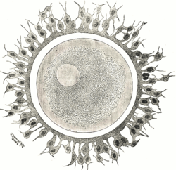
Zygote

 Clash Royale CLAN TAG#URR8PPP
| Zygote (cell) | |
|---|---|
 | |
| Details | |
| Days | 0 |
| Precursor | Gametes |
| Gives rise to | Morula |
| Identifiers | |
| MeSH | D015053 |
| TE | E2.0.1.2.0.0.9 |
Anatomical terminology [edit on Wikidata] | |
A zygote (from Greek ζυγωτός zygōtos "joined" or "yoked", from ζυγοῦν zygoun "to join" or "to yoke")[1] is a eukaryotic cell formed by a fertilization event between two gametes. The zygote's genome is a combination of the DNA in each gamete, and contains all of the genetic information necessary to form a new individual. In multicellular organisms, the zygote is the earliest developmental stage. In single-celled organisms, the zygote can divide asexually by mitosis to produce identical offspring.
Oscar Hertwig and Richard Hertwig made some of the first discoveries on animal zygote formation.
Contents
1 Fungi
2 Plants
3 Humans
4 In other species
5 In protozoa
6 See also
7 References
Fungi
In fungi, the sexual fusion of haploid cells is called karyogamy. The result of karyogamy is the formation of a diploid cell called zygote or zygospore. This cell may then enter meiosis or mitosis depending on the life cycle of the species.
Plants
In plants, the zygote may be polyploid if fertilization occurs between meiotically unreduced gametes.
In land plants, the zygote is formed within a chamber called the archegonium. In seedless plants, the archegonium is usually flask-shaped, with a long hollow neck through which the sperm cell enters. As the zygote divides and grows, it does so inside the archegonium.
Humans
Main articles: Development of the human body, Human fertilization
In human fertilization, a released ovum (a haploid secondary oocyte with replicate chromosome copies) and a haploid sperm cell (male gamete)—combine to form a single 2n diploid cell called the zygote. Once the single sperm enters the oocyte, it completes the division of the second meiosis forming a haploid daughter with only 23 chromosomes, almost all of the cytoplasm, and the sperm in its own pronucleus. The other product of meiosis is the second polar body with only chromosomes but no ability to replicate or survive. In the fertilized daughter, DNA is then replicated in the two separate pronuclei derived from the sperm and ovum, making the zygote's chromosome number temporarily 4n diploid. After approximately 30 hours from the time of fertilization, fusion of the pronuclei and immediate mitotic division produce two 2n diploid daughter cells called blastomeres.[2]
Between the stages of fertilization and implantation, the developing human is a preimplantation conceptus. There is some dispute about whether this conceptus should no longer be referred to as an embryo, but should now be referred to as a proembryo, which is terminology that traditionally has been used to refer to plant life. Some ethicist and legal scholars make the argument that it is incorrect to call the conceptus an embryo, because it will later differentiate into both intraembryonic and extraembryonic tissues,[3] and can even split to produce multiple embryos (identical twins). Others have pointed out that so-called extraembryonic tissues are really part of the embryo's body that are no longer used after birth (much as milk teeth fall out after childhood). Further, as the embryo splits to form identical twins – leaving the original tissues intact – new embryos are generated, in a process similar to that of cloning an adult human.[4] In the US the National Institutes of Health has determined that the traditional classification of pre-implantation embryo is still correct.[5]
After fertilization, the conceptus travels down the oviduct towards the uterus while continuing to divide[6]mitotically without actually increasing in size, in a process called cleavage.[7] After four divisions, the conceptus consists of 16 blastomeres, and it is known as the morula.[8] Through the processes of compaction, cell division, and blastulation, the conceptus takes the form of the blastocyst by the fifth day of development, just as it approaches the site of implantation.[9] When the blastocyst hatches from the zona pellucida, it can implant in the endometrial lining of the uterus and begin the embryonic stage of development.
The human zygote has been genetically edited in experiments designed to cure inherited diseases.[10]
In other species
A Chlamydomonas zygote contains chloroplast DNA (cpDNA) from both parents; such cells are generally rare, since normally cpDNA is inherited uniparentally from the mt+ mating type parent. These rare biparental zygotes allowed mapping of chloroplast genes by recombination.
In protozoa
In the amoeba, reproduction occurs by cell division of the parent cell: first the nucleus of the parent divides into two and then the cell membrane also cleaves, becoming two "daughter" Amoebae.
See also
- Proembryo
References
^ "English etymology of zygote". etymonline.com. Archived from the original on 2017-03-30..mw-parser-output cite.citationfont-style:inherit.mw-parser-output .citation qquotes:"""""""'""'".mw-parser-output .citation .cs1-lock-free abackground:url("//upload.wikimedia.org/wikipedia/commons/thumb/6/65/Lock-green.svg/9px-Lock-green.svg.png")no-repeat;background-position:right .1em center.mw-parser-output .citation .cs1-lock-limited a,.mw-parser-output .citation .cs1-lock-registration abackground:url("//upload.wikimedia.org/wikipedia/commons/thumb/d/d6/Lock-gray-alt-2.svg/9px-Lock-gray-alt-2.svg.png")no-repeat;background-position:right .1em center.mw-parser-output .citation .cs1-lock-subscription abackground:url("//upload.wikimedia.org/wikipedia/commons/thumb/a/aa/Lock-red-alt-2.svg/9px-Lock-red-alt-2.svg.png")no-repeat;background-position:right .1em center.mw-parser-output .cs1-subscription,.mw-parser-output .cs1-registrationcolor:#555.mw-parser-output .cs1-subscription span,.mw-parser-output .cs1-registration spanborder-bottom:1px dotted;cursor:help.mw-parser-output .cs1-ws-icon abackground:url("//upload.wikimedia.org/wikipedia/commons/thumb/4/4c/Wikisource-logo.svg/12px-Wikisource-logo.svg.png")no-repeat;background-position:right .1em center.mw-parser-output code.cs1-codecolor:inherit;background:inherit;border:inherit;padding:inherit.mw-parser-output .cs1-hidden-errordisplay:none;font-size:100%.mw-parser-output .cs1-visible-errorfont-size:100%.mw-parser-output .cs1-maintdisplay:none;color:#33aa33;margin-left:0.3em.mw-parser-output .cs1-subscription,.mw-parser-output .cs1-registration,.mw-parser-output .cs1-formatfont-size:95%.mw-parser-output .cs1-kern-left,.mw-parser-output .cs1-kern-wl-leftpadding-left:0.2em.mw-parser-output .cs1-kern-right,.mw-parser-output .cs1-kern-wl-rightpadding-right:0.2em
^ Blastomere Encyclopædia Britannica Archived 2013-09-28 at the Wayback Machine. Encyclopædia Britannica Online. Encyclopædia Britannica Inc., 2012. Web. 06 Feb. 2012.
^ Larsen's Human Embryology. 4th Ed. Page 4.
^ Condic, Maureen L. (14 April 2014). "Totipotency: What It Is And What It Is Not". Stem Cells and Development. 23: 796–812. doi:10.1089/scd.2013.0364. PMC 3991987. PMID 24368070.
^ "Archived copy" (PDF). Archived from the original (PDF) on 2009-01-30. Retrieved 2009-02-17.CS1 maint: Archived copy as title (link)
^ O’Reilly, Deirdre. "Fetal development Archived 2011-10-27 at the Wayback Machine". MedlinePlus Medical Encyclopedia (2007-10-19). Retrieved 2009-02-15.
^ Klossner, N. Jayne and Hatfield, Nancy. Introductory Maternity & Pediatric Nursing, p. 107 (Lippincott Williams & Wilkins, 2006).
^ Neas, John F. "Human Development" Archived July 22, 2011, at the Wayback Machine. Embryology Atlas
^ Blackburn, Susan. Maternal, Fetal, & Neonatal Physiology, p. 80 (Elsevier Health Sciences 2007).
^ Human zygote edited genetically Archived 2015-05-18 at the Wayback Machine
| Preceded by Oocyte + Sperm | Stages of human development Zygote | Succeeded by Embryo |