Adrenal cortex

 Clash Royale CLAN TAG#URR8PPP
 | Adrenal cortex | |
|---|---|
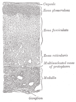
 Layers of cortex. | |
 The adrenal cortex | |
| Details | |
| Precursor | mesoderm[1] |
| Identifiers | |
| Latin | cortex glandulae suprarenalis |
| MeSH | D000302 |
| TA | A11.5.00.007 |
| FMA | 15632 |
Anatomical terminology [edit on Wikidata] | |
Situated along the perimeter of the adrenal gland, the adrenal cortex mediates the stress response through the production of mineralocorticoids and glucocorticoids, such as aldosterone and cortisol, respectively. It is also a secondary site of androgen synthesis.[2] Recent data suggest that adrenocortical cells under pathological as well as under physiological conditions show neuroendocrine properties; within the normal adrenal, this neuroendocrine differentiation seems to be restricted to cells of the zona glomerulosa and might be important for an autocrine regulation of adrenocortical function.[3]
Contents
1 Layers
2 Hormone synthesis
3 Production
3.1 Mineralocorticoids
3.2 Glucocorticoids
3.3 Androgens
4 Pathology
5 See also
6 References
7 External links
Layers
The adrenal cortex comprises three main zones, or layers. This anatomic zonation can be appreciated at the microscopic level, where each zone can be recognized and distinguished from one another based on structural and anatomic characteristics.[4] The adrenal cortex exhibits functional zonation as well: by virtue of the characteristic enzymes present in each zone, the zones produce and secrete distinct hormones.[4]
- Zona glomerulosa
- The outermost layer, the zona glomerulosa is the main site for production of aldosterone, a mineralocorticoid, by the action of the enzyme aldosterone synthase (also known as CYP11B2).[5][6] Aldosterone is largely responsible for the long-term regulation of blood pressure.[7]Aldosterone's effects are on the distal convoluted tubule and collecting duct of the kidney where it causes increased reabsorption of sodium and increased excretion of both potassium (by principal cells) and hydrogen ions (by intercalated cells of the collecting duct).[7] Sodium retention is also a response of the distal colon, and sweat glands to aldosterone receptor stimulation. Although sustained production of aldosterone requires persistent calcium entry through low-voltage activated Ca2+ channels, isolated zona glomerulosa cells are considered nonexcitable, with recorded membrane voltages that are too hyperpolarized to permit Ca2+ channels entry.[8] However, mouse zona glomerulosa cells within adrenal slices spontaneously generate membrane potential oscillations of low periodicity; this innate electrical excitability of zona glomerulosa cells provides a platform for the production of a recurrent Ca2+ channels signal that can be controlled by angiotensin II and extracellular potassium, the 2 major regulators of aldosterone production.[8] Angiotensin II originates from plasmatic angiotensin I after the conversion of angiotensinogen by renin produced by the juxtaglomerular cells of the kidney.[9]
- The expression of neuron-specific proteins in the zona glomerulosa cells of human adrenocortical tissues has been predicted and reported by several authors[3][10][11] and it was suggested that the expression of proteins like the neuronal cell adhesion molecule (NCAM) in the cells of the zona glomerulosa reflects the regenerative feature of these cells, which would lose NCAM immunoreactivity after moving to the zona fasciculata.[3][12] However, together with other data on neuroendocrine properties of zona glomerulosa cells, NCAM expression may reflect a neuroendocrine differentiation of these cells.[3]
- Zona fasciculata
- Situated between the glomerulosa and reticularis, the zona fasciculata is responsible for producing glucocorticoids, such as 11-deoxycorticosterone, corticosterone, and cortisol in humans. Cortisol is the main glucocorticoid under normal conditions and its actions include mobilization of fats, proteins, and carbohydrates, but it does not increase under starvation conditions.[9] Additionally, cortisol enhances the activity of other hormones including glucagon and catecholamines. The zona fasciculata secretes a basal level of cortisol but can also produce bursts of the hormone in response to adrenocorticotropic hormone (ACTH) from the anterior pituitary.
- Zona reticularis
- The inner most cortical layer, the zona reticularis produces androgens, mainly dehydroepiandrosterone (DHEA), DHEA sulfate (DHEA-S), and androstenedione (the precursor to testosterone) in humans.[9]
Hormone synthesis

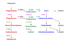
Adrenal steroid pathways
All adrenocortical steroid hormones are synthesized from cholesterol. Cholesterol is transported into the adrenal gland. The steps up to this point occur in many steroid-producing tissues. Subsequent steps to generate aldosterone and cortisol, however, primarily occur in the adrenal cortex:
- Progesterone → (hydroxylation at C21) → 11-Deoxycorticosterone → (two further hydroxylations at C11 and C18) → Aldosterone
- Progesterone → (hydroxylation at C17) → 17-alpha-hydroxyprogesterone → (hydroxylation at C21) → 11-Deoxycortisol → (hydroxylation at C11) → Cortisol

Adrenal steroid hormone synthesis steps
Production
The adrenal cortex produces a number of different corticosteroid hormones.
Mineralocorticoids
The primary mineralocorticoid, aldosterone, is produced in the adrenocortical zona glomerulosa by the action of the enzyme aldosterone synthase (also known as CYP11B2).[5][6] Aldosterone is largely responsible for the long-term regulation of blood pressure.[7] Aldosterone effects on the distal convoluted tubule and collecting duct of the kidney where it causes increased reabsorption of sodium and increased excretion of both potassium (by principal cells) and hydrogen ions (by intercalated cells of the collecting duct).[7] Sodium retention is also a response of the distal colon, and sweat glands to aldosterone receptor stimulation. Although sustained production of aldosterone requires persistent calcium entry through low-voltage activated Ca2+ channels, isolated zona glomerulosa cells are considered nonexcitable, with recorded membrane voltages that are too hyperpolarized to permit Ca2+ channels entry.[8] However, mouse zona glomerulosa cells within adrenal slices spontaneously generate membrane potential oscillations of low periodicity; this innate electrical excitability of zona glomerulosa cells provides a platform for the production of a recurrent Ca2+ channels signal that can be controlled by angiotensin II and extracellular potassium, the 2 major regulators of aldosterone production.[8] Angiotensin II originates from plasmatic angiotensin I after the conversion of angiotensinogen by renin produced by the juxtaglomerular cells of the kidney.[9]
Glucocorticoids
They are produced in the zona fasciculata. The primary glucocorticoid released by the adrenal gland is cortisol in humans and corticosterone in many other animals. Its secretion is regulated by the hormone ACTH from the anterior pituitary. Upon binding to its target, cortisol enhances metabolism in several ways:
- It stimulates the release of amino acids from the body
- It stimulates lipolysis, the breakdown of fat
- It stimulates gluconeogenesis, the production of glucose from newly released amino acids and lipids
- It increases blood glucose levels in response to stress, by inhibiting glucose uptake into muscle and fat cells
- It strengthens cardiac muscle contractions
- It increases water retention
- It has anti-inflammatory and anti-allergic effects
Androgens
They are produced in the zona reticularis. The most important androgens include:
Testosterone: a hormone with a wide variety of effects, ranging from enhancing muscle mass and stimulation of cell growth to the development of the secondary sex characteristics.
Dihydrotestosterone (DHT): a metabolite of testosterone, and a more potent androgen than testosterone in that it binds more strongly to androgen receptors.
Androstenedione (Andro): an androgenic steroid produced by the testes, adrenal cortex, and ovaries. While androstenediones are converted metabolically to testosterone and other androgens, they are also the parent structure of estrone.
Dehydroepiandrosterone (DHEA): It is the primary precursor of natural estrogens. DHEA is also called dehydroisoandrosterone or dehydroandrosterone. The reticularis also produces DHEA-sulfate due to the actions of a sulfotransferase, SULT2A1.[13]
Pathology
Adrenal insufficiency (e.g. due to Addison's disease)- Cushing's syndrome
- Conn's syndrome
See also
- Adrenarche
- Adrenopause
References
^ "Embryology of the adrenal gland". Retrieved 2007-12-11..mw-parser-output cite.citationfont-style:inherit.mw-parser-output qquotes:"""""""'""'".mw-parser-output code.cs1-codecolor:inherit;background:inherit;border:inherit;padding:inherit.mw-parser-output .cs1-lock-free abackground:url("//upload.wikimedia.org/wikipedia/commons/thumb/6/65/Lock-green.svg/9px-Lock-green.svg.png")no-repeat;background-position:right .1em center.mw-parser-output .cs1-lock-limited a,.mw-parser-output .cs1-lock-registration abackground:url("//upload.wikimedia.org/wikipedia/commons/thumb/d/d6/Lock-gray-alt-2.svg/9px-Lock-gray-alt-2.svg.png")no-repeat;background-position:right .1em center.mw-parser-output .cs1-lock-subscription abackground:url("//upload.wikimedia.org/wikipedia/commons/thumb/a/aa/Lock-red-alt-2.svg/9px-Lock-red-alt-2.svg.png")no-repeat;background-position:right .1em center.mw-parser-output .cs1-subscription,.mw-parser-output .cs1-registrationcolor:#555.mw-parser-output .cs1-subscription span,.mw-parser-output .cs1-registration spanborder-bottom:1px dotted;cursor:help.mw-parser-output .cs1-hidden-errordisplay:none;font-size:100%.mw-parser-output .cs1-visible-errorfont-size:100%.mw-parser-output .cs1-subscription,.mw-parser-output .cs1-registration,.mw-parser-output .cs1-formatfont-size:95%.mw-parser-output .cs1-kern-left,.mw-parser-output .cs1-kern-wl-leftpadding-left:0.2em.mw-parser-output .cs1-kern-right,.mw-parser-output .cs1-kern-wl-rightpadding-right:0.2em
^ Anatomy Atlases - Microscopic Anatomy, plate 15.292 – "Adrenal Gland"
^ abcd Ehrhart-Bornstein M, Hilbers U (1998). "Neuroendocrine properties of adrenocortical cells". Horm. Metab. Res. 30 (6–7): 436–9. doi:10.1055/s-2007-978911. PMID 9694576.
^ ab Whitehead, Saffron A.; Nussey, Stephen (2001). Endocrinology: an integrated approach. Oxford: BIOS. p. 122. ISBN 1-85996-252-1.
^ ab Curnow KM, Tusie-Luna MT, Pascoe L, et al. (October 1991). "The product of the CYP11B2 gene is required for aldosterone biosynthesis in the human adrenal cortex". Mol. Endocrinol. 5 (10): 1513–22. doi:10.1210/mend-5-10-1513. PMID 1775135.
^ ab Zhou M, Gomez-Sanchez CE (July 1993). "Cloning and expression of a rat cytochrome P-450 11 beta-hydroxylase/aldosterone synthase (CYP11B2) cDNA variant". Biochem. Biophys. Res. Commun. 194 (1): 112–7. doi:10.1006/bbrc.1993.1792. PMID 8333830.
^ abcd Marieb Human Anatomy & Physiology 9th edition, chapter:16, page:629, question number:14
^ abcd Hu C, Rusin CG, Tan Z, Guagliardo NA, Barrett PQ (June 2012). "Zona glomerulosa cells of the mouse adrenal cortex are intrinsic electrical oscillators". J. Clin. Invest. 122 (6): 2046–53. doi:10.1172/JCI61996. PMC 3966877. PMID 22546854.
^ abcd Dunn R. B.; Kudrath W.; Passo S.S.; Wilson L.B. (2011). "10". Kaplan USMLE Step 1 Physiology Lecture Notes. pp. 263–289.
^ Lefebvre H, Cartier D, Duparc C, et al. (March 2002). "Characterization of serotonin(4) receptors in adrenocortical aldosterone-producing adenomas: in vivo and in vitro studies". J. Clin. Endocrinol. Metab. 87 (3): 1211–6. doi:10.1210/jcem.87.3.8327. PMID 11889190.
^ Ye P, Mariniello B, Mantero F, Shibata H, Rainey WE (October 2007). "G-protein-coupled receptors in aldosterone-producing adenomas: a potential cause of hyperaldosteronism". J. Endocrinol. 195 (1): 39–48. doi:10.1677/JOE-07-0037. PMID 17911395.
^ Haidan A, Bornstein SR, Glasow A, Uhlmann K, Lübke C, Ehrhart-Bornstein M (February 1998). "Basal steroidogenic activity of adrenocortical cells is increased 10-fold by coculture with chromaffin cells". Endocrinology. 139 (2): 772–80. doi:10.1210/endo.139.2.5740. PMID 9449652.
^ Rainey WE, Nakamura Y (February 2008). "Regulation of the adrenal androgen biosynthesis". J. Steroid Biochem. Mol. Biol. 108 (3–5): 281–6. doi:10.1016/j.jsbmb.2007.09.015. PMC 2699571. PMID 17945481.
External links
Anatomy photo:40:04-0203 at the SUNY Downstate Medical Center – "Posterior Abdominal Wall: Blood Supply to the Suprarenal Glands"
MedicalMnemonics.com: 180 2201 412
Histology image: 14502loa – Histology Learning System at Boston University