Egg cell

 Clash Royale CLAN TAG#URR8PPP
 | Egg cell | |
|---|---|
 A human ovum with corona radiata surrounding it | |
Anatomical terminology [edit on Wikidata] |

Human Ovum Cell
The egg cell, or ovum (plural ova), is the female reproductive cell (gamete) in oogamous organisms. The egg cell is typically not capable of active movement, and it is much larger (visible to the naked eye) than the motile sperm cells. When egg and sperm fuse, a diploid cell (the zygote) is formed, which rapidly grows into a new organism.
Contents
1 History
2 Animals
2.1 Human and mammal ova
2.2 Ooplasm
2.3 Ova development in oviparous animals
2.4 Ovoviviparity
3 Plants
4 Other organisms
5 See also
6 References
7 External links
History
While the non-mammalian animal egg was obvious, the doctrine ex ovo omne vivum ("every living [animal comes from] an egg"), associated with William Harvey (1578–1657), was a rejection of spontaneous generation and preformationism as well as a bold assumption that mammals also reproduced via eggs. Karl Ernst von Baer discovered the mammalian ovum in 1827, and Edgar Allen discovered the human ovum in 1928. The fusion of spermatozoa with ova (of a starfish) was observed by Oskar Hertwig in 1876.[1][2]
Animals
In animals, egg cells are also known as ova (singular ovum, from the Latin word ovum meaning egg or egg cell). The term ovule in animals is used for the young ovum of an animal. In vertebrates, ova are produced by female gonads (sexual glands) called ovaries. A number of ova are present at birth in mammals and mature via oogenesis. White et al. disproved the longstanding dogma that all of the ova are produced before birth. The team from the Vincent Center for Reproductive Biology, Massachusetts, Boston showed that oocyte formation takes place in ovaries of reproductive-age women.[3][4][clarification needed] This report challenged a fundamental belief, held since the 1950s, that female mammals are born with a finite supply of eggs that is depleted throughout life and exhausted at menopause.[5]
Human and mammal ova

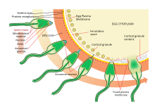
Diagram of a human egg cell
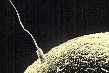
Ovum and sperm fusing together
The process of fertilizing an ovum (Top to bottom)
In all mammals the ovum is fertilized inside the female body.
The human ova grow from primitive germ cells that are embedded in the substance of the ovaries. Each of them divides repeatedly to give secretions of the uterine glands, ultimately forming a blastocyst.[6]
The ovum is one of the largest cells in the human body, typically visible to the naked eye without the aid of a microscope or other magnification device. The human ovum measures approximately 0.1 mm in diameter.[7][8]
Ooplasm
Ooplasm (also: oöplasm) is the yolk of the ovum, a cell substance at its center, which contains its nucleus, named the germinal vesicle, and the nucleolus, called the germinal spot.[9]
The ooplasm consists of the cytoplasm of the ordinary animal cell with its spongioplasm and hyaloplasm, often called the formative yolk; and the nutritive yolk or deutoplasm, made of rounded granules of fatty and albuminoid substances imbedded in the cytoplasm.[9]
Mammalian ova contain only a tiny amount of the nutritive yolk, for nourishing the embryo in the early stages of its development only. In contrast, bird eggs contain enough to supply the chick with nutriment throughout the whole period of incubation.[9]
Ova development in oviparous animals
In the oviparous animals (all birds, most fish, amphibians and reptiles) the ova develop protective layers and pass through the oviduct to the outside of the body. They are fertilized by male sperm either inside the female body (as in birds), or outside (as in many fish). After fertilization, an embryo develops, nourished by nutrients contained in the egg. It then hatches from the egg, outside the mother's body. See egg for a discussion of eggs of oviparous animals.
The egg cell's cytoplasm and mitochondria are the sole means the egg is able to reproduce by mitosis and eventually form a blastocyst after fertilization.
Ovoviviparity
There is an intermediate form, the ovoviviparous animals: the embryo develops within and is nourished by an egg as in the oviparous case, but then it hatches inside the mother's body shortly before birth, or just after the egg leaves the mother's body. Some fish, reptiles and many invertebrates use this technique.
Plants
Nearly all land plants have alternating diploid and haploid generations. Gametes are produced by the gametophyte, which is the haploid generation. The female gametophyte produces structures called archegonia, and the egg cells form within them via mitosis. The typical bryophyte archegonium consists of a long neck with a wider base containing the egg cell. Upon maturation, the neck opens to allow sperm cells to swim into the archegonium and fertilize the egg. The resulting zygote then gives rise to an embryo, which will grow into a new diploid individual (sporophyte). In seed plants, a structure called ovule, which contains the female gametophyte. The gametophyte produces an egg cell. After fertilization, the ovule develops into a seed containing the embryo.[10]
In flowering plants, the female gametophyte (sometimes referred to as the embryo sac) has been reduced to just eight cells inside the ovule. The gametophyte cell closest to the micropyle opening of the ovule develops into the egg cell. Upon pollination, a pollen tube delivers sperm into the gametophyte and one sperm nucleus fuses with the egg nucleus. The resulting zygote develops into an embryo inside the ovule. The ovule in turn develops into a seed and in many cases the plant ovary develops into a fruit to facilitate the dispersal of the seeds. Upon germination, the embryo grows into a seedling.[10]
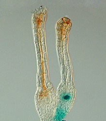
Gene expression pattern determined by histochemical GUS assays in Physcomitrella patens. The Polycomb gene FIE is expressed (blue) in unfertilised egg cells of the moss Physcomitrella patens (right) and expression ceases after fertilisation in the developing diploid sporophyte (left). In situ GUS staining of two female sex organs (archegonia) of a transgenic plant expressing a translational fusion of FIE-uidA under control of the native FIE promoter
In the moss Physcomitrella patens, the Polycomb protein FIE is expressed in the unfertilised egg cell (Figure, right) as the blue colour after GUS staining reveals. Soon after fertilisation the FIE gene is inactivated (the blue colour is no longer visible, left) in the young embryo.
[11]
Other organisms
In algae, the egg cell is often called oosphere.[citation needed]Drosophila oocytes develop in individual egg chambers that are supported by nurse cells and surrounded by somatic follicle cells. The nurse cells are large polyploid cells that synthesize and transfer RNA, proteins and organelles to the oocytes. This transfer is followed by the programmed cell death (apoptosis) of the nurse cells. During the course of oogenesis, 15 nurse cells die for every oocyte that is produced.[12] In addition to this developmentally regulated cell death, egg cells may also undergo apoptosis in response to starvation and other insults.[12]
See also
- Egg
- Fertilization
- Insemination
- Menstrual cycle
- Ova bank
- Ovulation
- Polar body
- Pollination
- Pregnancy
- Spawn (biology)
References
^ Needham, Joseph (1959). A History of Embryology (2d revised ed.). Cambridge: Cambridge University Press..mw-parser-output cite.citationfont-style:inherit.mw-parser-output qquotes:"""""""'""'".mw-parser-output code.cs1-codecolor:inherit;background:inherit;border:inherit;padding:inherit.mw-parser-output .cs1-lock-free abackground:url("//upload.wikimedia.org/wikipedia/commons/thumb/6/65/Lock-green.svg/9px-Lock-green.svg.png")no-repeat;background-position:right .1em center.mw-parser-output .cs1-lock-limited a,.mw-parser-output .cs1-lock-registration abackground:url("//upload.wikimedia.org/wikipedia/commons/thumb/d/d6/Lock-gray-alt-2.svg/9px-Lock-gray-alt-2.svg.png")no-repeat;background-position:right .1em center.mw-parser-output .cs1-lock-subscription abackground:url("//upload.wikimedia.org/wikipedia/commons/thumb/a/aa/Lock-red-alt-2.svg/9px-Lock-red-alt-2.svg.png")no-repeat;background-position:right .1em center.mw-parser-output .cs1-subscription,.mw-parser-output .cs1-registrationcolor:#555.mw-parser-output .cs1-subscription span,.mw-parser-output .cs1-registration spanborder-bottom:1px dotted;cursor:help.mw-parser-output .cs1-hidden-errordisplay:none;font-size:100%.mw-parser-output .cs1-visible-errorfont-size:100%.mw-parser-output .cs1-subscription,.mw-parser-output .cs1-registration,.mw-parser-output .cs1-formatfont-size:95%.mw-parser-output .cs1-kern-left,.mw-parser-output .cs1-kern-wl-leftpadding-left:0.2em.mw-parser-output .cs1-kern-right,.mw-parser-output .cs1-kern-wl-rightpadding-right:0.2em
^ Lopata, Alex (April 2009). "History of the Egg in Embryology". Journal of Mammalian Ova Research. 26 (1): 2–9. doi:10.1274/jmor.26.2.
^ White, Yvonne A R (26 February 2012). "Oocyte formation by mitotically active germ cells purified from ovaries of reproductive-age women". Nature Medicine (18): 413–421. doi:10.1038/nm.2669. PMC 3296965. Retrieved 28 March 2017.
^ Woods, Dori C (18 April 2013). "Isolation, characterization and propagation of mitotically active germ cells from adult mouse and human ovaries". Nature Protocols (8): 966–988. doi:10.1038/nprot.2013.047. Retrieved 28 March 2017.
^ Massachusetts General Hospital. "Egg-producing stem cells isolated from adult human ovaries". ScienceDaily. Retrieved 28 March 2017.
^ Regan, Carmen L. (2001). "Pregnancy". In Worell, Judith. Encyclopedia of Women and Gender: Sex Similarities and Differences and the Impact of Society on Gender, Volume 1. Academic Press. p. 859. ISBN 9780122272455. Retrieved 2013-11-03.
^ Search result of "120 micrometers" in Level O Biology – Google books
^ Alberts, Bruce; Johnson, Alexander; Lewis, Julian; Raff, Martin; Roberts, Keith; Walter, Peter (2002). "Eggs".
^ abc "The Ovum". Gray's Anatomy. Retrieved 2010-10-18.
^ ab Esau, K. (1977). Anatomy of seed plants (second ed.). New York: John Wiley and Sons. ISBN 978-0-471-24520-9.
^ Mosquna, Assaf; Katz, Aviva; Decker, Eva L.; Rensing, Stefan A.; Reski, Ralf; Ohad, Nir (2009). "Regulation of stem cell maintenance by the Polycomb protein FIE has been conserved during land plant evolution". Development. 136: 2433–2444. doi:10.1242/10.1242/dev.035048.
^ ab McCall K (October 2004). "Eggs over easy: cell death in the Drosophila ovary". Dev. Biol. 274 (1): 3–14. doi:10.1016/j.ydbio.2004.07.017. PMID 15355784.
External links
The Ovarian Kaleidoscope Database description of 1800 genes involved in ovarian functions
Sex | |
|---|---|
| Biological terms |
|
| Sexual reproduction |
|
| Sexuality |
|
| |
Authority control |
|
|---|